Upper Back Back Bones Diagram - Anatomy Of The Back Spine And Back Muscles Kenhub / Place right elbow on left elbow.. Other causes of upper back pain include: Discover (and save!) your own pins on pinterest Human backbone diagram, bone, human backbone diagram. Both the deltoid and the trapezius are firmly attached to … See lumbar spine anatomy diagram stock video clips.
The lumbar and sacrum region make up the bone of the lower back anatomy. See lumbar spine anatomy diagram stock video clips. The body is the largest part of the vertebrae and the part that bears the most weight. For more anatomy content please follow us and visit our website: Lumbar spine anatomy diagram images.
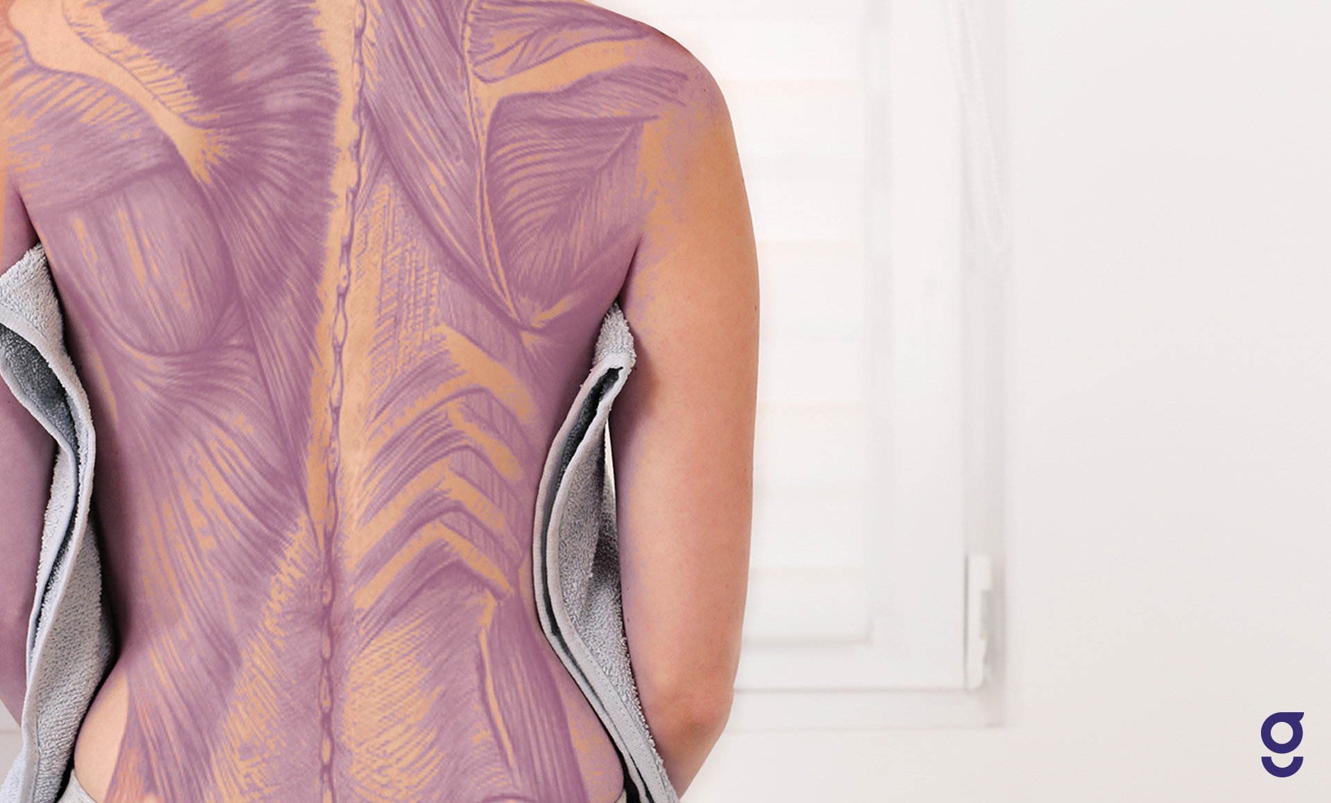
Back Muscles Anatomy Of Upper Middle Lower Back Pain In Diagrams Goodpath from images.ctfassets.net Thoracic region of the spine. Each vertebra consists of the following parts: Discover (and save!) your own pins on pinterest It runs from the neck to the upper back. Straining a muscle or ligament your back. The human spine is composed of 4 sections of vertebrae. Your lower back contains 5 vertebral bones stacked above each other with intervertebral discs in between. Lack of strength in the muscles of your back (for example, from not doing much exercise) sitting at a computer for long periods of time.
There are 12 bones that make up the upper back, which doctors call the thoracic spine.
Your lower back contains 5 vertebral bones stacked above each other with intervertebral discs in between. The bones of the chest and upper back combine to form the strong, protective rib cage around the vital thoracic organs such as the heart and lungs. Our latest youtube film is ready to run. The third thoracic vertebrae is a small vertebra in the upper middle back that plays an integral role in supporting the rib cage. The four principal types of bones are long, short, flat and irregular. Other muscles are small and cover much less space. So many patients receive diagnostic imaging reports full of terms and anatomical locations which are unknown and mysterious to them. The spine diagram shown below, consists of many bones or vertebrae,soft discs,the spinal cord, and spinal nerves. The cervical spine protects the nerves connecting to the brain, allowing the head to move freely while supporting its weight. It consists of seven vertebrae. It runs from the neck to the upper back. The bones of the pelvis and lower back work together to support the body's weight, anchor the abdominal and hip muscles, and protect the delicate vital organs of the vertebral and abdominopelvic cavities. The thoracic spine starts beneath the neck and is comprised of 12 vertebrae, labeled t1 through t12, which go down the back of the torso (figure 1).unlike the cervical spine and lumbar spine, the thoracic spine is relatively immobile because each of its vertebrae are connected to a pair of ribs (one on.
Stand up with your arms on the side of your body. The lumbar and sacrum region make up the bone of the lower back anatomy. Lack of strength in the muscles of your back (for example, from not doing much exercise) sitting at a computer for long periods of time. The bones of the pelvis and lower back work together to support the body's weight, anchor the abdominal and hip muscles, and protect the delicate vital organs of the vertebral and abdominopelvic cavities. For optimum maximum muscle contraction, squeeze the shoulder blades together at the end of each pull, before releasing back to the front.
Thoracic Spine from www.spineuniverse.com Discover (and save!) your own pins on pinterest Studying a spine diagram is one way to better understand many of the individual components of the back bone and how they might relate to a symptomatic back, neck or sciatica pain condition. Back of right upper extremity. The teres majo r muscles work with the rotator cuff muscles to stabilize. Thoracic region of the spine. The rib cage also anchors the bones of the head, neck, shoulders, and arms to the trunk of the body. Other muscles are small and cover much less space. The bones of the chest and upper back combine to form the strong, protective rib cage around the vital thoracic organs such as the heart and lungs.
The third thoracic vertebrae is a small vertebra in the upper middle back that plays an integral role in supporting the rib cage.
Studying a spine diagram is one way to better understand many of the individual components of the back bone and how they might relate to a symptomatic back, neck or sciatica pain condition. However, irritation of the large back and. Back muscles anatomy here include the trapezius, latissimus dorsi, rhomboid and levator scapulae. Lack of strength in the muscles of your back (for example, from not doing much exercise) sitting at a computer for long periods of time. The cervical spine protects the nerves connecting to the brain, allowing the head to move freely while supporting its weight. It consists of seven vertebrae. The spine diagram shown below, consists of many bones or vertebrae,soft discs,the spinal cord, and spinal nerves. The lumbar and sacrum region make up the bone of the lower back anatomy. This is my video about the muscles of the back. For optimum maximum muscle contraction, squeeze the shoulder blades together at the end of each pull, before releasing back to the front. Back of skull (occipital bone) fused vertebrae (5) (sacrum) hand bones (metacarpals) finger bones. The back functions are many, such as to house and protect the spinal cord, hold the body and head upright, and adjust the movements of the upper and lower limbs. It is like that for several reasons, all of which you can understand by looking at the anatomy of the thoracic spine.
Bones, discs, and joints in your lower back. The four principal types of bones are long, short, flat and irregular. The lumbar spine connects to the thoracic spine above and the hips below. The third thoracic vertebrae is a small vertebra in the upper middle back that plays an integral role in supporting the rib cage. The human spine is composed of 4 sections of vertebrae.
Anatomy Of The Upper Back from www.spineuniverse.com Back muscles anatomy here include the trapezius, latissimus dorsi, rhomboid and levator scapulae. Back of right upper extremity. Powerful muscles that move the head and arms attach to these bones as well. Each vertebra consists of the following parts: License image the deltoid, teres major, teres minor, infraspinatus, supraspinatus (not shown) and subscapularis muscles (not shown) all extend from the scapula to the humerus and act on the shoulder joint. The bones of the chest and upper back combine to form the strong, protective rib cage around the vital thoracic organs such as the heart and lungs. Powerful muscles that move the head and arms attach to these bones as well. It comprises the vertebral column (spine) and two compartments of back muscles;
Discover (and save!) your own pins on pinterest
This is my video about the muscles of the back. Stand up with your arms on the side of your body. There are 12 bones that make up the upper back, which doctors call the thoracic spine. Anatomy upper limb bones and cartilages bones of upper limb. Back muscles anatomy here include the trapezius, latissimus dorsi, rhomboid and levator scapulae. It is like that for several reasons, all of which you can understand by looking at the anatomy of the thoracic spine. The upper back is a complex area containing a number of muscles that perform various actions on the scapulae (shoulder blades) and humerus. It comprises the vertebral column (spine) and two compartments of back muscles; The body is the largest part of the vertebrae and the part that bears the most weight. Bones, discs, and joints in your lower back. Straining a muscle or ligament your back. The bones of the chest and upper back combine to form the strong, protective rib cage around the vital thoracic organs such as the heart and lungs. The back's muscles start at the top of the back (named the cervical vertebrae) and go to the tailbone (also named the coccyx).
For optimum maximum muscle contraction, squeeze the shoulder blades together at the end of each pull, before releasing back to the front back bones diagram. The third thoracic vertebrae is a small vertebra in the upper middle back that plays an integral role in supporting the rib cage.


0 Komentar